Atopic Dermatitis: Overview of Current Diagnosis
- No clear consensus has emerged regarding the diagnostic work-up that should be performed when evaluating patients with AD, particularly in adult patients. Diagnostic approaches vary widely.1
- Chronic pruritus/itching induces scratch behavior. While this can serve as a physiologic self-protective mechanism to prevent the body from being hurt by harmful external agents, it also damages skin and increases inflammation. This further exacerbates pruritus and often leads to the ‘‘itch-scratch cycle.’’2
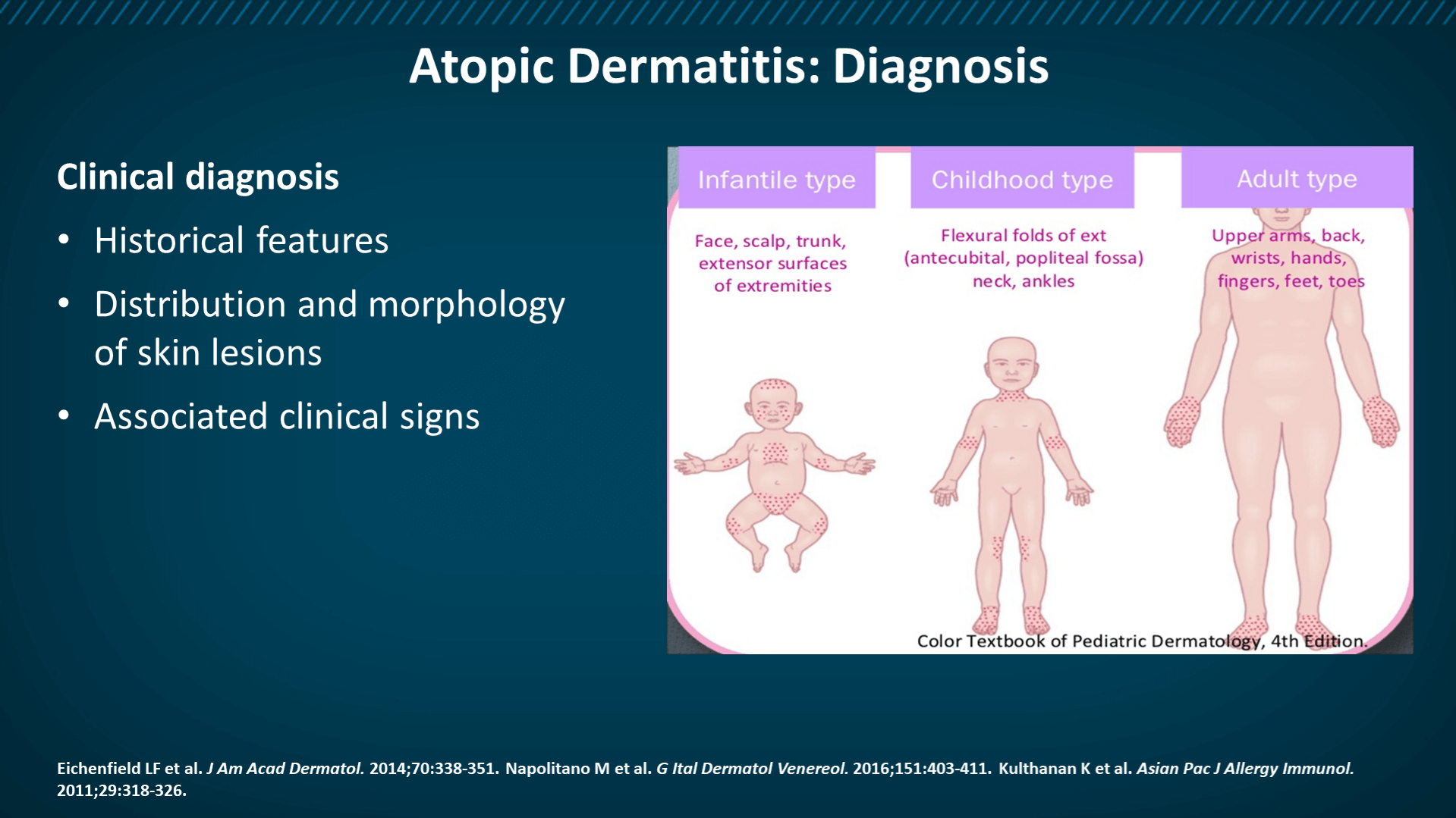
- Clinical phenotypes and endophenotypes are characterized by a wide range of heterogeneity in the onset, course, and presentation of AD, as well as in individual comorbidities.
- The diagnosis of AD remains clinical because there are no known reliable biomarkers that can distinguish AD from other diseases.3,4
- Guidelines for the management of AD issued in 2014 reported 28 different scales for the measurement of disease severity, without a single gold standard emerging.4
The following symptoms and features are also commonly seen in AD:
- Pruritus
- Chronic and relapsing course
- Early age of onset
- Peripheral eosinophilia
- Immunoglobulin (Ig) E reactivity
- Staphylococcus aureus superinfection
- Personal history of asthma or hay fever
- History of atopic diseases in a first-degree relative1
Diagnosis: American Academy of Dermatology
According to the American Academy of Dermatology,2,3 patients with presumed AD should have their diagnosis made based on a combination of features.
| Essential Features2,3 (must be present) | Important Features2,3 (support diagnosis; seen in most cases) |
· Pruritus *Patterns include: | · Early age of onset · Atopy · Personal and/or family history · IgE reactivity · Xerosis |
“Associated features” help suggest the diagnosis of AD but are nonspecific.2,3 These include:
- Atypical vascular responses
- Keratosis pilaris, pityriasis alba, hyperlinear palms, and ichthyosis
- Ocular/periorbital changes
- Perifollicular accentuation, lichenification, and prurigo lesions
In addition, other conditions must be excluded, such as scabies, seborrheic dermatitis, contact dermatitis, ichthyosis, cutaneous T-cell lymphoma, psoriasis, photosensitivity dermatoses, immunodeficiency diseases, and erythroderma of other causes.2,3
For additional information on the diagnosis of AD, we encourage you to visit the following references as well as our “Additional Reading” sections.
References
- Kim BS. Atopic dermatitis. Updated June 3, 2020 (https://emedicine.medscape.com/article/1049085-overview).
- American Academy of Neurology. Atopic dermatitis clinical guideline (https://www.aad.org/practicecenter/quality/clinical-guidelines/atopic-dermatitis/diagnosis-and-assessment).
- Eichenfield LF, et al. Guidelines of care for the management of atopic dermatitis: section 1. Diagnosis and assessment of atopic dermatitis. J Am Acad Dermatol. 2014;70:338-351.